Anatomy of the Ear
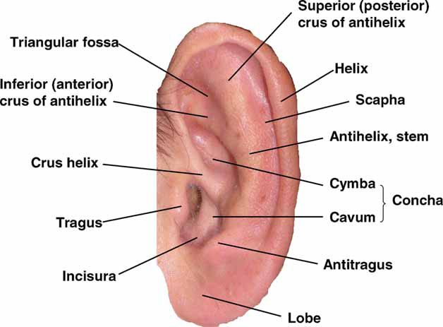
The anatomy of the external ear, also known as the auricle or pinna, is complex [Hunter and Yotsuyanagi, [2005]] and remarkably inaccurately described by most authors. The major landmarks of the external ear are depicted in Figure 1. The external ear consists of skin (with adnexa), cartilage, and six intrinsic muscles. The anatomy of the various components of the ear are described below, and illustrations are shown each time in the section describing the various features of the components.

Figure 1
- Antihelix:
- A Y-shaped curved cartilaginous ridge arising from the antitragus and separating the concha, triangular fossa, and scapha. The antihelix represents a folding of the conchal cartilage and it usually has similar prominence to a well-developed helix. The stem (the part below the bifurcation) of the normal antihelix is gently curved and branches about two thirds of the way along its course to form the broad fold of the superior (posterior) antihelical crus, and the more sharply folded inferior (anterior) crus. The inferior and superior crura of the antihelix can vary both in volume and degree of folding.
- Antihelix, Inferior Crus:
- The lower cartilaginous ridge arising at the bifurcation of the antihelix that ends beneath the fold of the ascending helix, and separates the concha from the triangular fossa. The inferior antihelical crus runs in an anterior and slightly superior direction, is usually sharply defined, and appears less variable than its superior counterpart. A synonym is anterior crus of the antihelix.
- Antihelix, Superior Crus:
- The upper cartilaginous ridge arising at the bifurcation of the antihelix that separates the scapha from the triangular fossa. The superior crus runs in a superior and slightly anterior direction and is usually less sharply folded than the lower portion and inferior crus. A synonym is the posterior crus of the antihelix.
- Antitragus:
- The anterosuperior cartilaginous protrusion lying between the incisura and the origin of the antihelix. The anterosuperior margin of the antitragus forms the posterior wall of the incisura.
- Concha:
- The fossa bounded by the tragus, incisura, antitragus, antihelix, inferior crus of the antihelix, and root of the helix, into which opens the external auditory canal. It is usually bisected by the crus helix into the cymba superiorly and cavum inferiorly.
- Frankfurt Horizontal:
- A plane connecting the lowest point on the lower margin of each orbit and highest point on the upper margin of the external auditory meatus [Farkas, [1981]]. The Frankfurt horizontal or Frankfurt plane is used as the general horizontal plane of the head and as reference point for other planes and structures.
- Helix:
- The outer rim of the ear that extends from the superior insertion of the ear on the scalp (root) to the termination of the cartilage at the earlobe. The helix can be divided into three approximate parts: the ascending helix, which extends vertically from the root; the superior helix, which begins at the top of the ascending portion, extends horizontally and curves posteriorly to the site of Darwin tubercle (vide infra); the descending helix (sometimes called posterior), which begins inferior to Darwin tubercle and extends to the superior border of the earlobe. The lower portion of the posterior part is often non-cartilaginous. The border of the helix usually forms a rolled rim but the helix is highly variable in shape. Lange [1966] developed a graphic classification of folding variants.
- Helix, Crus:
- The continuation of the anteroinferior ascending helix, which extends in a posteroinferior direction into the cavity of the concha above the external auditory meatus (Figure. 1). The average crus helix extends about one half to two thirds the distance across the concha. A synonym is crista helicis.
- Lobe:
- The soft, fleshy, inferior part of the pinna. It is bounded on its posterosuperior border by the end of the descending helix, on the anterosuperior border by the inferior border of the antitragus and superiorly by the incisura. The earlobe is highly variable in size and in the degree of attachment of the anteroinferior portion to the face.
- Scapha:
- The groove between the helix and the antihelix.
- Tragus:
- A posterior, slightly inferior, protrusion of skin-covered cartilage, anterior to the auditory meatus. The inferoposterior margin of the tragus forms the anterior wall of the incisura.
- Triangular Fossa:
- The concavity bounded by the superior and inferior crura of the antihelix and the ascending portion of the helix.
Anatomical Variation
Anomalies of the ear include quantitative traits and qualitative features of the entire ear, and of the individual components.
- Variation in size (macrotia; microtia; anotia).
- Variation in position (low-set ears; posterior angulation of the ear).
- Variations of the individual anatomical parts: antihelix; antitragus; concha; helix; lobe; scapha; tragus; triangular fossa.
- Named ear anomalies (crumpled ear; cryptotia; cupped ear; lop ear; preauricular and auricular pits; preauricular and auricular tags; preauricular ectopias; prominent ear; question mark ear; detachment of ascending helix; satyr ear; shell ear; Stahl ear).