Anatomy of the Face and Cranium
Head shape and upper face shape are closely related to the shape of the bony skull. Figures 1 and 2 show the bony anatomy of the face. Many anthropological landmarks, bony and soft tissue, are illustrated in Figures 3 and 4.

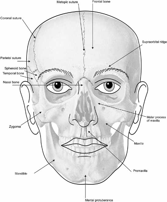
Figure 1
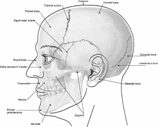
Figure 2
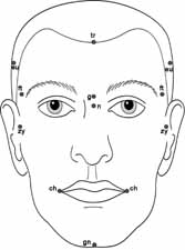
Figure 3
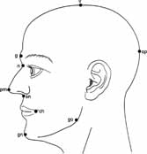
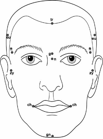
Figure 4
The anatomy of the various structures is described in more detail below.
- Cranium:
- The upper part of the skull consists of paired frontal and parietal bones and a single posterior occipital bone (Figures 1 and 2). In early life these bones are separated by five major sutures (Figures 1 and 2). Three, the coronal, lambdoidal and squamosal, are paired, and two, the sagittal and metopic, are single. Cranial growth normally occurs perpendicular to each of these major sutures.
- Forehead:
- The part of the face above the eyebrows, below the hairline and between the temples. The paired frontalis muscles join in the midline and adhere to the superficial fascia over the frontal bone. These muscles effect forehead wrinkling or furrowing. They have no bony attachments, but inferiorly the fibres blend with the muscles encircling the eyelids. From these attachments the fibers are directed upward, and join the galea aponeurotica below the coronal suture. The galea aponeurotica is a layer of dense fibrous tissue which covers the upper part of the cranium and attaches posteriorly to the occipital bone. It is closely connected to the integument by the firm, dense, fibro-fatty layer which forms the superficial fascia of the scalp. It cannot be wrinkled or furrowed because it does not contain muscle fibres. The anterior hairline is typically situated at the junction of frontalis muscle and galea aponeurotica.
- Glabella:
- The most prominent point on the frontal bone above the root of the nose.
- Supra-orbital Ridge:
- The supraorbital portion of the frontal bones.
- Midface:
- This is a region and not an anatomical term. It extends, superiorly, from the inferior orbital margin to, inferiorly, the level of nasal base. It is formed by the maxilla (upper jaw) and zygoma. Traditionally, the nose and premaxilla are not included in the midface.
- Maxilla:
- These paired bones form, by their union, the upper jaw and contain the upper dentition. Each assists in forming the boundaries of three cavities - the palate, floor and lateral wall of the nose (frontal or malar process), and floor of the orbit. Each bone consists of a body and four processes - zygomatic, malar (frontal), alveolar and palatine.
- Malar Process (Synonym: Frontal Process):
- The most medial and superior part of the maxilla. It forms the medial border of the inferior bony orbit, and is contiguous with the lateral boundary of the nasal bridge.
- Zygoma:
- The part of the temporal bone of the skull that forms the prominence of the cheek. It is also known as the zygomatic bone or arch, the malar bone (creating confusion with the malar process of the maxilla), the cheek bone and the yoke bone. The zygomatic arch is composed of the malar process of the maxilla, medially, the zygoma, centrally, and the temporal bone, posterolaterally. It forms part of the part of the lateral wall and floor of the orbit.
- Premaxilla:
- The part of the maxilla in which the 4 upper incisors develop, which forms the primary palate, and underlies the philtrum and upper lip.
- Lower Face:
- The part of the face between the mouth and the inferior point of the chin.
- Cheek:
- The soft tissues between the zygoma and mandible.
- Mandible:
- The lower jaw in which the lower teeth reside. It consists of a curved, horizontal portion, the body, and two perpendicular portions, the rami, which unite with the ends of the body nearly at right angles.
- Chin:
- The inferior portion of the face lying inferior to the lower lip and including the central prominence of the lower jaw.
- Neck:
- The part of the body connecting the head with the shoulders.