Anatomy of the Periorbital Region
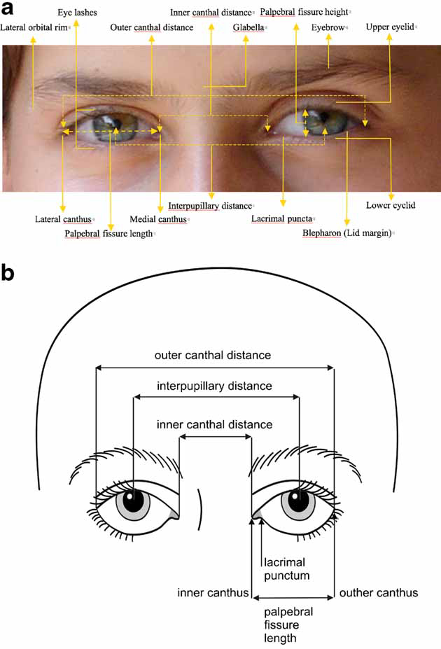
The general anatomy of the non-globe periorbital region is depicted in Figure 1. The definitions for the terms utilized in describing the features within this region are listed alphabetically. The anatomy of the various structures is described in more detail below.
Figure 1
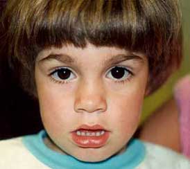
Figure 2
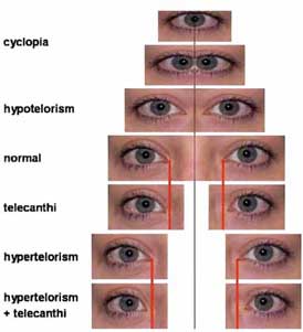
Figure 3
- Brow:
- The soft tissue at the junction of the frontalis and orbicularis oculi muscles, overlying the bony supraorbital ridge.
- Eyebrow:
- The arch of hair on the brow (Fig. 2) [Goss, [1959]]. The eyebrows usually extend further laterally than medially, in relation to the eye, and are wider and thicker medially. Based on observed localized abnormalities of the eyebrow, it is useful to divide the eyebrow into three parts: medial, middle (central), and lateral. The hairs of the medial part are oriented laterally, while those of the middle (central) part are oriented superolaterally. The transition between the middle and lateral parts is less frequently visible. Some syndromes have unique patterns of aberrations in one or more of these three areas. The eyebrow is sometimes referred to as the supercilium.
- Eye Spacing:
- There is wide variation in interorbital distance and in the placement of the canthi [Cohen et al., [1995]]. A number of terms in this article address the nomenclature of these variations. Several of the terms are commonly confused (especially telecanthus and hypertelorism). Some of the variations are illustrated in Figure 3.
- Eyelashes:
- Hairs that emanate from the margins of the eyelids [Goss, [1959]].
- Eyelid (Synonym: Blepharon, palpebra {plural: palpebrae}):
- A fold of skin and its subcutaneous components that covers the anterior globe. The upper lid is bounded by the soft tissue overlying the inferior border of the bony supraorbital ridge and inferiorly by the lid margin. The lower lid is bounded by the soft tissue overlying the infraorbital rim and superiorly by the lid margin. A crescent-shaped crease on the upper eyelid represents the location of attachment of the levator palpebrae muscle to the orbicularis oculi muscle [Goss, [1959]].
- Lacrimal Punctum {Plural: puncta}:
- This structure represents the external aperture of the tear duct system. It can be absent, malpositioned, or obstructed [Ogawa and Gonnering, [1991]], and several terms below address these findings.
Note that the plurality of the terms is variable. The default chosen is to specify the singular form of the term unless the term relates to a pair of structures and only makes sense in the plural form (e.g., Eyes, closely spaced) or refers to a structure with many elements (e.g., Eyelashes, sparse). The plurality of the terms was ignored when they were alphabetized and the terms were grouped together (e.g., Eye and Eyes are grouped together, and not interrupted by Eyelashes).